About C2 Learn 2023
CALM Summer School for Light Microscopy
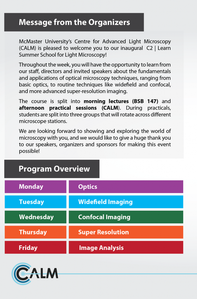
• Practical course on fundamentals and applications of optical microscopy
• Theory and application lectures in the morning, hands-on sessions in the afternoon
• Expecting a minimum of 50 participants (with practical sessions capped at 20
participants), both internal and external to McMaster University
• Opportunity to meet decision makers like principal investigators, facility directors
and managers
• Train the future generation of microscopists and help build a microscopy community
at McMaster University and Southern Ontario at large
• Opportunity to showcase latest equipment through demos, workshops, and talks
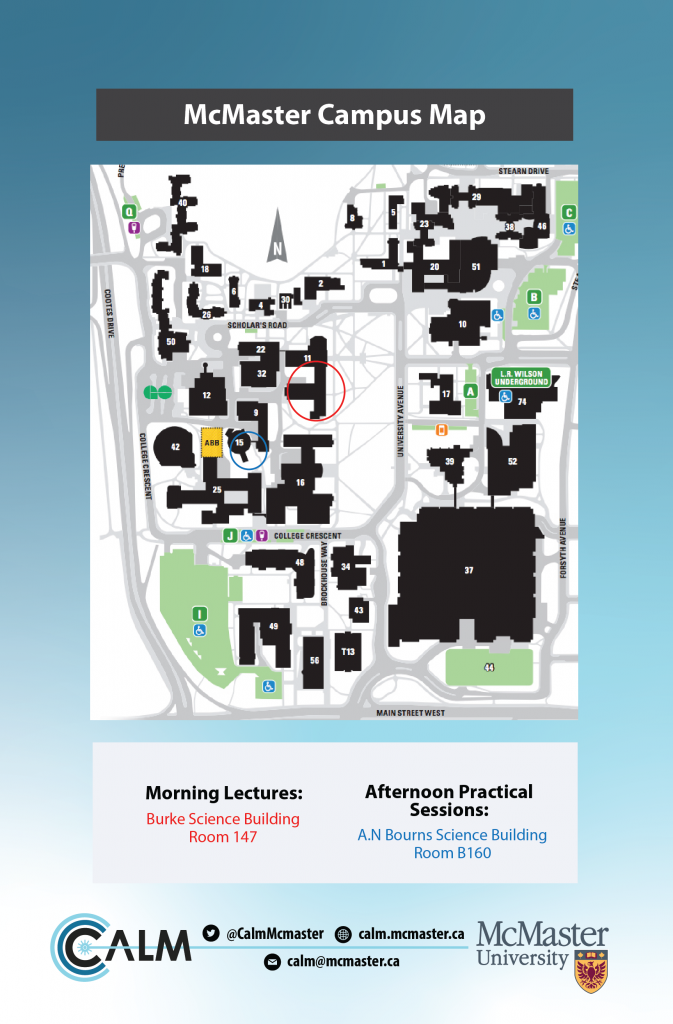
Location
Lectures (morning) – Burke Science Building room 147
Practicals (afternoon) – Center for Advanced Light Microscopy (CALM) – Arthur Bourns Building room B160
Course Dates
June 19 – 23 2023
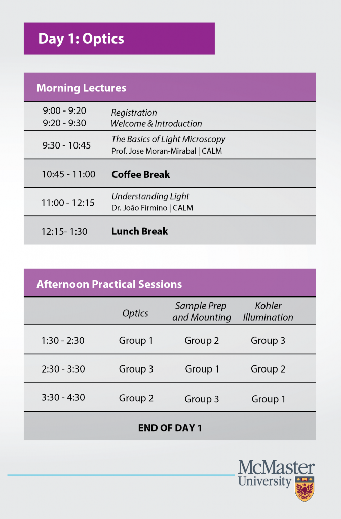
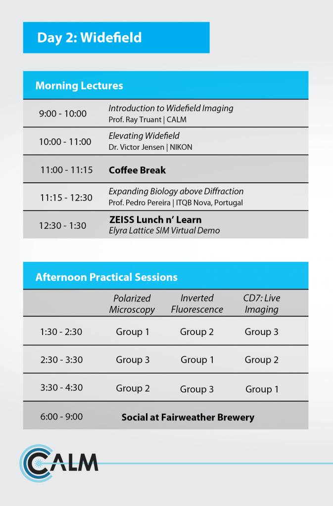
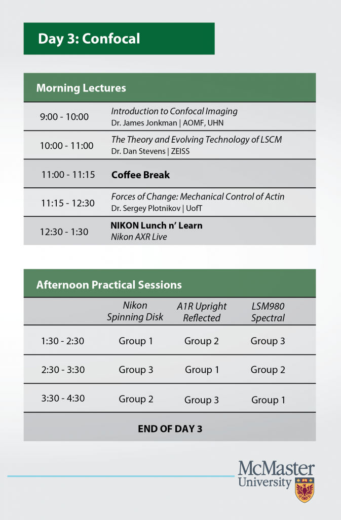
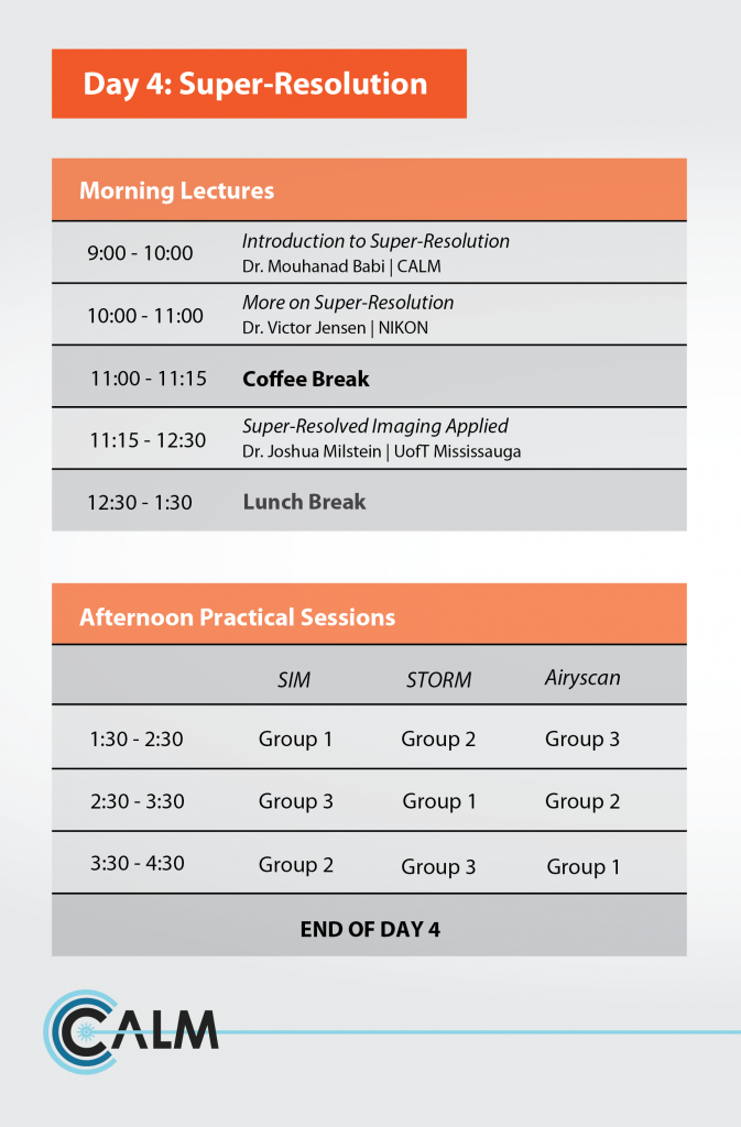
Program
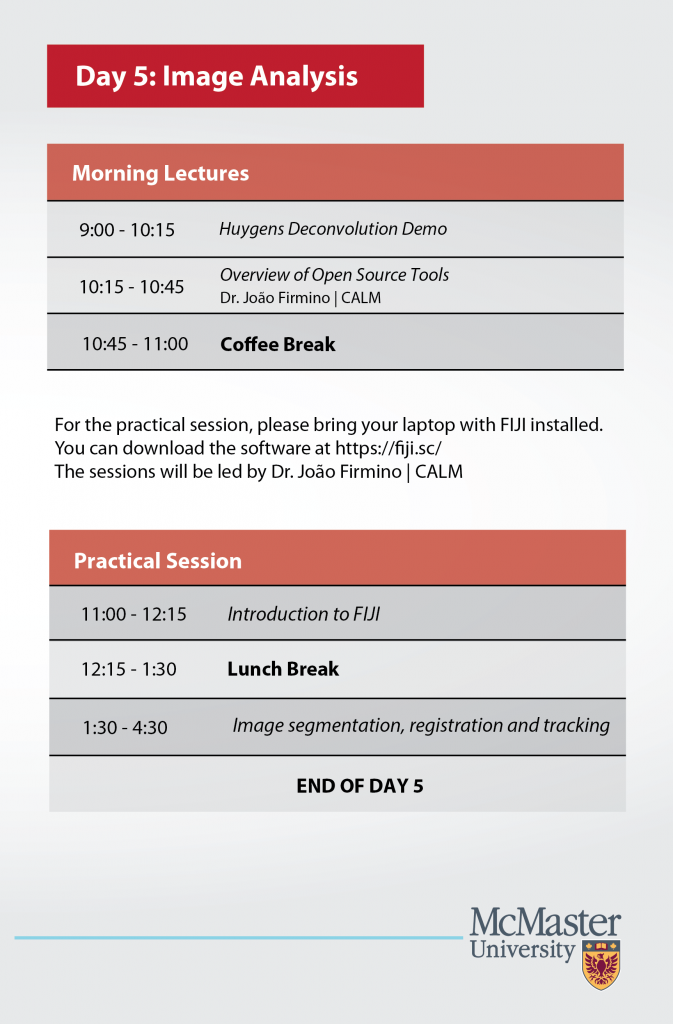
For Huygens Deconvolution demo, please download the appropriate package here: http://gofile.me/6Z5S7/SL5qyTFiM
We will share a temporary demo license and additional sample data files to download in due time.
Registration
Practical registration is now closed. Only theoretical sessions available at the moment!
| I WANT TO LEARN ABOUT MICROSCOPY! |
|---|
Invited Speakers
Prof. Pedro Pereira
Instituto de Tecnologia Química e Biológica António Xavier in Lisbon
https://www.itqb.unl.pt/research/biology/intracellular-microbial-infection-biology
Prof. Pereira is a cell biologist with experience in microbiology, host-pathogen interactions, and advanced microscopy approaches. He graduated in applied chemistry from NOVA university in Lisbon in 2006 and completed a master’s in biotechnology the following year. He then finished his PhD in cell biology in 2013, where he explored questions related to antibiotic resistance and cell division in the human pathogen S. aureus. During this time, he started to explore different microscopy approaches such as classical fluorescence microscopy, super-resolution microscopy, and atomic force microscopy. This interest prompted his move to University College in London, UK, where he did a postdoc focusing on the development of hardware, software, and probe-based technological innovations for (super-resolution) microscopy (e.g., NanoJ-Fluidics). In 2019 he returned to Portugal, where, since October 2021 he has his own research group at ITQB NOVA, the Intracellular Microbial Infection Biology (IMIB) laboratory. Pedro is currently interested in understanding how extracellular bacterial pathogens transition into an intracellular infection “lifestyle” and how antibiotic challenges modulate this behaviour.
Prof. Sergey Plotnikov
University of Toronto
Dr. Sergey Plotnikov is an Assistant Professor in the Department of Cell and Systems Biology. He completed his Ph.D. at the Institute of Marine Biology, Vladivostok, Russia. The Plotnikov Lab aims to uncover how migration of animal cells is controlled by mechanical signals form the extracellular environment. The lab uses a multifaceted experimental approach, combining cell biology, molecular biology, and biophysics techniques with high-resolution quantitative optical microscopy to address the following specific questions: (i) unraveling molecular mechanisms that transduce physical signals experienced by cells into activity of signaling pathways, (ii) assess contribution of directed cell migration guided by physical cues to pathological conditions, (iii) understanding how mechanosensitive cellular responses are modulated by chemical guidance cues, such as spatial gradients of growth factors.
Prof. Joshua Milstein
University of Toronto Mississauga
https://www.utm.utoronto.ca/milsteinlab/
The Milstein laboratory for biological physics focuses on studying the spatial organization and dynamical motion of DNA, which, while being intimately involved in genetic function, has operational effects that are not clearly understood. Errors in genetic expression, besides arising from mistakes in the code itself, could also manifest by macromolecules that over-twist or over-stretch the DNA, problems in packing within the cell or nucleus, long-range interference between genes, and so on. Our research aims to clarify important biological questions from how bacteria transform into harmful pathogens to how embryonic stem cells differentiate. To address these questions, we build and develop single-molecule tools such as optical tweezers, for applying controlled forces to biological molecules, and super-resolution microscopes, for imaging fine structure within bacteria and the cell nucleus. Our laboratory is a truly-interdisciplinary environment filled with a range of researchers, from physicists and biomedical engineers to biologists and computer scientists.
James Jonkman
University Health Network in Toronto
James is Manager of the Advanced Optical Microscopy Facility (AOMF). He has developed and currently manages the largest microscopy facility in Canada. With over 40 instruments at 4 sites, he and his team at the AOMF help nearly 500 users per year with
every manner of optical microscopy, from basic fluorescence and brightfield acquisition to advanced techniques such as Super-resolution, FRAP, FRET, and 3D analysis. James is particularly passionate about teaching and training users. He has been running week-long optical microscopy courses in the AOMF for 14 years, has co-authored several book chapters and review articles, presented at numerous conferences and webinars, and has helped as an organizer and instructor for the Montreal Light Microscopy Course (MLMC). James was also the co-president of the Canadian Cytometry and Microscopy Association (CCMA) from 2014-2017.
We would like to thank our sponsors:

![]()
![]()
![]()